7.2 Alternative Secondary Structures of DNA
7.2.1 Left-handed (i.e., “Z”) DNA
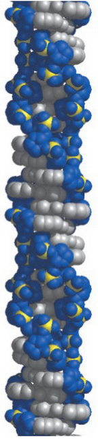
Figure 7.7: Z DNA
Polynucleotides with alternating purines and pyrimidines (i.e., GC, GT, AC, or AT repeats) in each strand are able to form left-handed DNA.
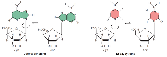
Figure 7.8: Rotation of Bases Around the N-glycosidic Bond
The rotation of bases around the N-glycosidic bond in figure 7.8 permits various geometric relationships of the base to the sugar.
The syn and anti forms of left-handed DNA refer to the 180ο opposite conformations of the bases to the sugar.
Left-handed DNA requires an anti conformation for pyrimidines and a syn conformation for purines.
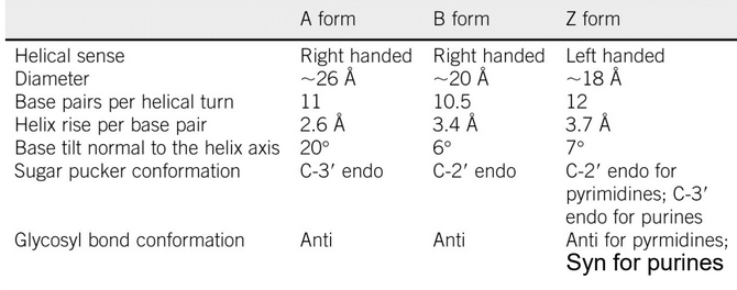
Figure 7.9: Comparisons Between Different Types of DNA
Figure 7.9 compares the parameters and features of A form, B form, and Z form DNA. The function of Z DNA in vivo appears to be related to gene regulations and relieving conformational stress.
7.2.2 Hairpins
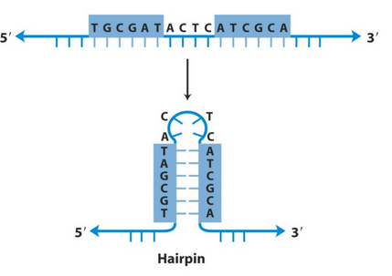
Figure 7.10: Hairpins
In single-stranded nucleic acid molecules, self-complementary sequence elements allow the polymer to fold back onto itself and form structures called hairpins.
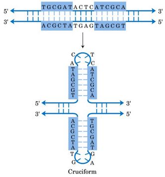
Figure 7.11: Cruciforms
A palindromic DNA sequence is a segment of duplex DNA where its bases exhibit a twofold rotational symmetry about the axis. Double hairpins (see figure 7.11), often called cruciform structures, can be formed with palindromic DNA sequences.
Note that many restriction endonucleases also have their palindromic sequnces as their cognate recognition sites.
7.2.3 Triple helices and Hoogsteen pairs
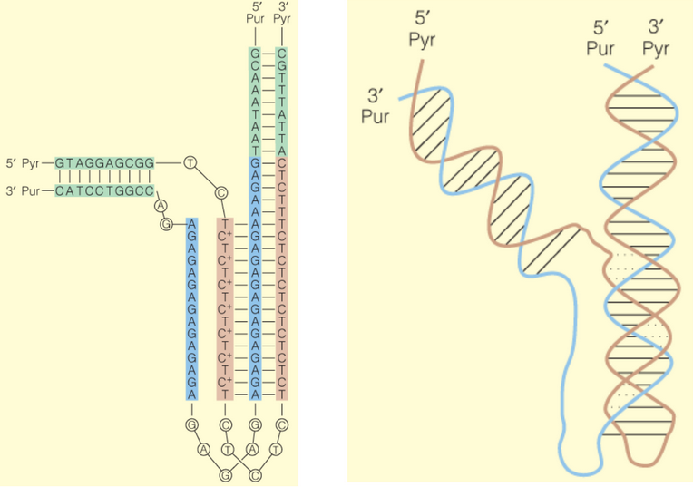
Figure 7.12: Schematic of a Triple Helix and Hoogsteen Pairs
A triple helix (see figure 7.12) is a molecule that has one all-pyrimidine containing strand and one all-purine containing strand.
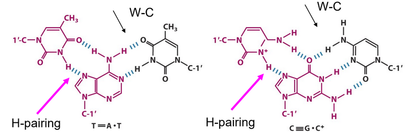
Figure 7.13: Watson-Crick and Hoogsteen Type Pairings
The formation of triple helices involve normal Watson-Crick base pairings and the Hoogsteen-type pairing (see 7.13).
Hoogsteen interactions occur on the major groove side of A or G.
7.2.4 Quadruplex DNA
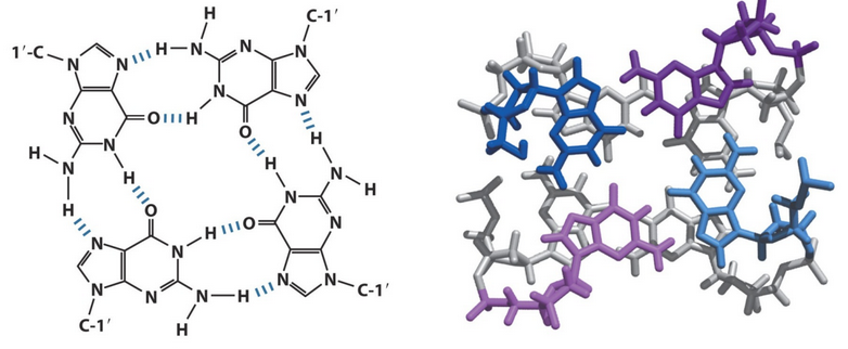
Figure 7.14: Guanosine Tetraplexes
Guanine-rich repeats (see figure 7.14) can form tetraplexes.
7.2.5 Circular DNA and supercoiling
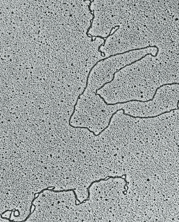
Figure 7.15: Circular DNA
Circular DNA molecules do not have free 5’ or 3’ ends. Instead, these molecules may involve a single strand or two intertwined strands (in a double helix).
Supercoiling is a tertiary structural feature of DNA that involves the higher-order folding of elements in regular, secondary DNA structure.
Supercoiled DNA have twists of the DNA double helix itself - the helix axis cross over itself at least one time.
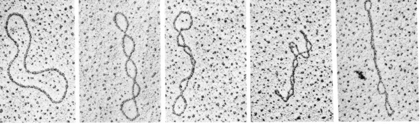
Figure 7.16: Topoisomers in Different Degrees of Supercoiling
A topoisomer is a strand of circular DNA that can assume relaxed and supercoiled forms. Topoisomers only differ in their topologies (hence the term “topoisomer”).
Figure 7.16 displays several topoisomers with increasing degrees of supercoiling from left to right.
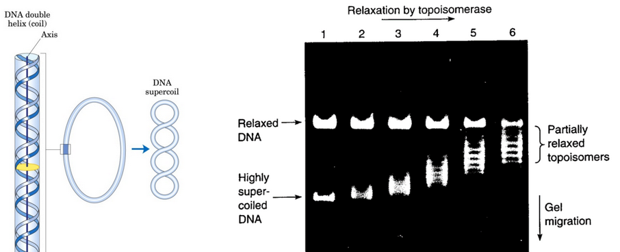
Figure 7.17: Topoisomerases in Action
A topoisomerase is an enzyme that is able to cut and reseal DNA. Because of this, topoisomers can be interconverted via cutting and resealing DNA (see figure 7.17).