2.6 Analysis of Amino Acids in Proteins
Several (self-explanatory) graphics (taken off prof. Davey’s slides) are presented in this section:
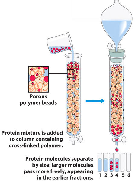
Figure 2.25: A Size-Exclusion Column Chromatography
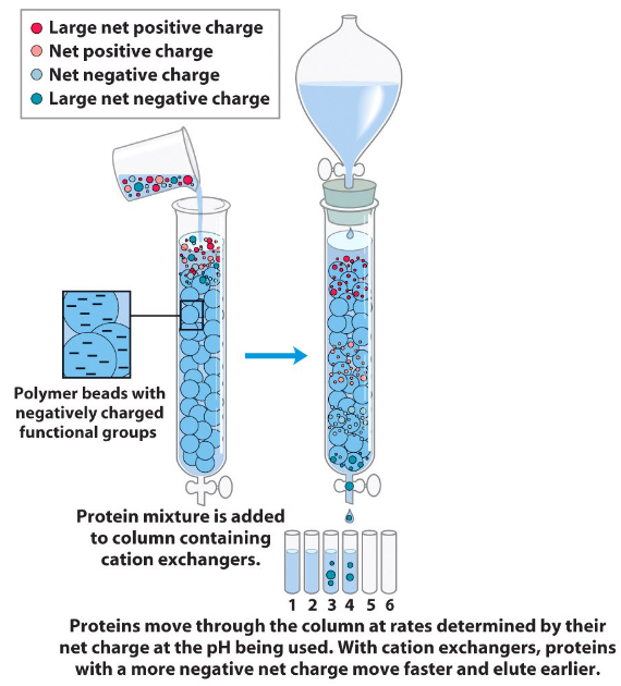
Figure 2.26: An Ion-Exchange Column Chromatography
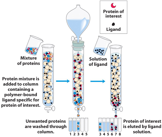
Figure 2.27: An Affinity Column Chromatography
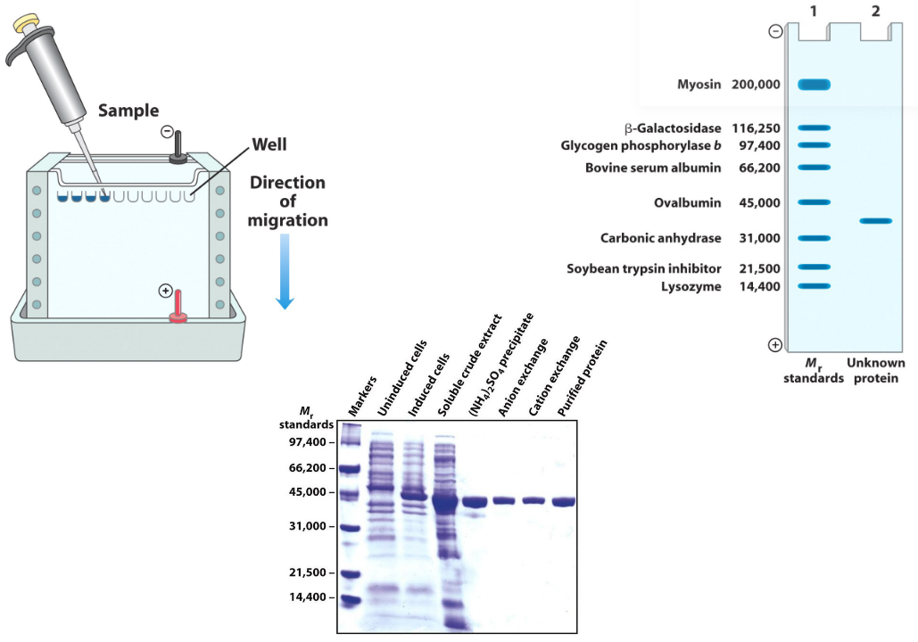
Figure 2.28: A SDS-PAGE Gel
Figure 2.28 displays a sodium dodecyl sulfate-polyacrylamide (SDS-PAGE) gel electrophoresis setup. Here, proteins are separated based on their molecular weights.
In an SDS-PAGE gel electrophoresis, proteins are first denatured; the denatured protein samples will then run towards the cathode as the samples are surrounded by negatively-charged SDS molecules.
An image of an actual SDS-PAGE gel is shown on the bottom of figure 2.28.