13.1 Background Information for Performing MRI
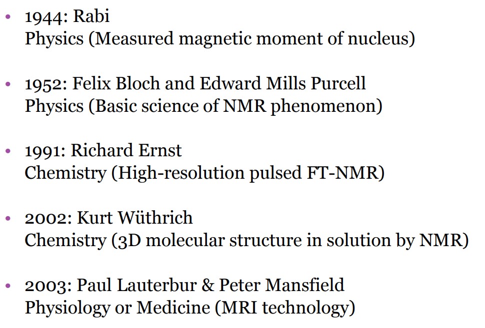
Figure 13.2: Nobel Prizes Won in Magnetic Resonance
There are many kinds of MRI, including the following:
- Nuclear Magnetic Resonance (i.e., NMR)
- Magnetic Resonance Imaging (i.e., MRI)
- Echo-Planar Imaging (i.e., EPI)
- functional MRI (i.e., fMRI)
- Magnetic Resonance Spectroscopy (i.e., MRS)
- MR Spectroscopic Imaging (i.e., MRSI)
13.1.1 Hydrogen Atoms
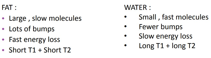
The human body is generally made up of fat and water, both of which have many atoms - 63% of the human body is made up of hydrogen atoms, and these atoms each have an NMR signal.
Consequently, MRI uses hydrogen because it only has one proton and easily aligns with the MRI magnet. This is because the hydrogen atom has a property called “spin” - this, in essence, produces a small magnetic field and will cause the nucleus to produce an NMR signal.
13.1.2 Magnetic Principles
Spinning hydrogen protons are like small, weak magnets that align with an external magnetic field. These protons spin about the axis of theexternal magnetic field at the resonance frequency.
There is also a slight excess of protons that are aligned with the field. The number of protons that are aligned with the field is so large that classical mechanics can be utilized.
n=γBo
The above is the formula for MRI frequency, where γ is the gyromagnetic ratio.
If an electromagnetic RF pulse is applied at the resonance frequency, then the protons can absorb the energy and jump to a higher energy state. On a macro level, the magnetization vector spirals down towards the XY plane.
Once the RF transmitter is turned off, three things happen at the same time:
- The absorbed RF energy is retransmitted
- Excited spins begin to return to the original Mz orientation
- Protons begin to dephase (i.e., T2 and T2* relaxation)
13.1.3 Basic Physics of MRI
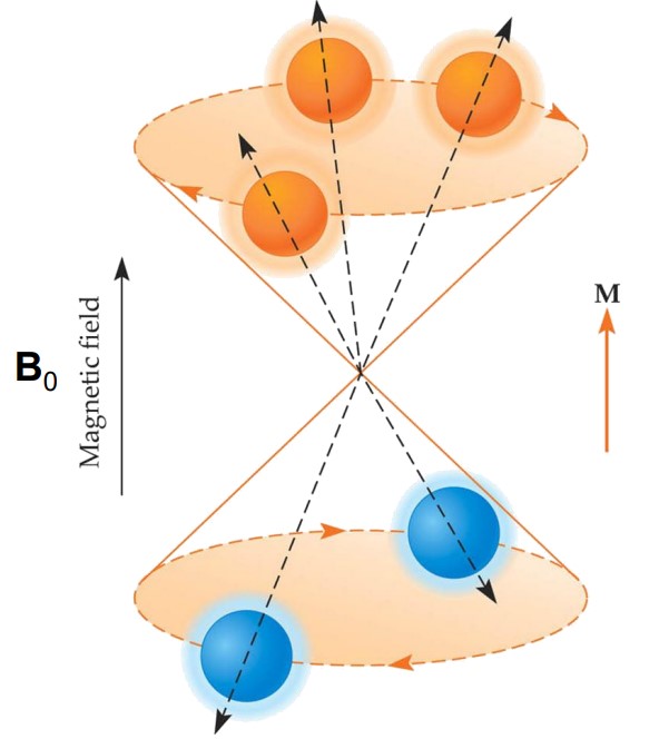
Figure 13.3: Illustration of a Magnetic Field
In the above figure M is parallel ot B0 as transverse components of magnetic moments are randomly oriented.
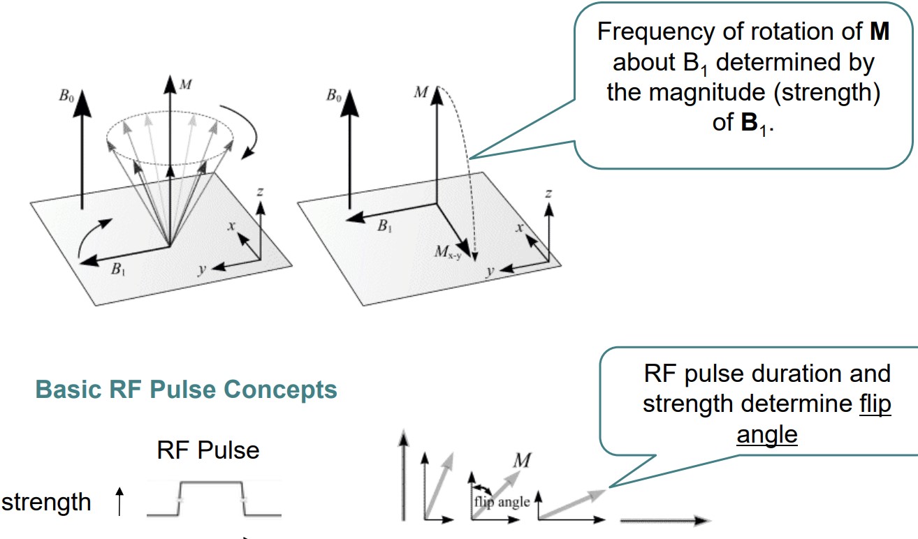
Figure 13.4: Illustration of Concepts in MRI
M is the difference between the amount of protons on the top level versus the amount of protons on the bottom (i.e., the anti-parallel state).
The proton density is the number of parallel states per unit volume - this also affects a sample’s signal-producing capabilities.