9.1 SEM and TEM
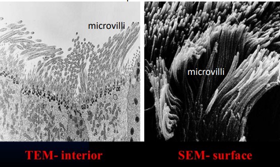
Figure 9.1: Epithelial Cells Viewed Using SEM and TEM
In a Scanning Electron Microscopy (i.e., SEM), electrons that are deflected by samples are of interest. These deflections provide useful information about the kind of object (e.g., cells, tissues, and even entire organisms) being dealt with. There are also different SEM techniques for large-volume imaging (e.g., Serial Blockface SEM).
In a Transmission Electron Microscopy (i.e., TEM), electrons that are transmitted through the object are of interest. This also implies that cell and tissue samples must be cut into thin slices (i.e., about 30 - 60 nm). Because of this, TEM is limited by the area and the thickness of samples - it does, however, provide a higher resolution than SEM.