10.1 Missing Wedge Problem and Electron Tomography
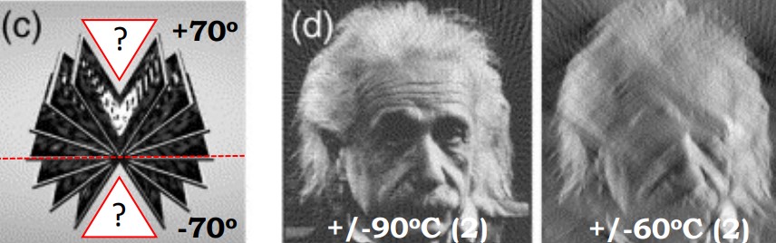
Figure 10.3: Illustration of the Missing Wedge Problem
Specimens cannot be viewed at angles beyond 60 to 70 degrees, so certain views may be inaccessible. Consequently, anisotropic resolution may result: features may appear elongated (i.e., smeared) in the electron beam’s direction (i.e., the z-axis).
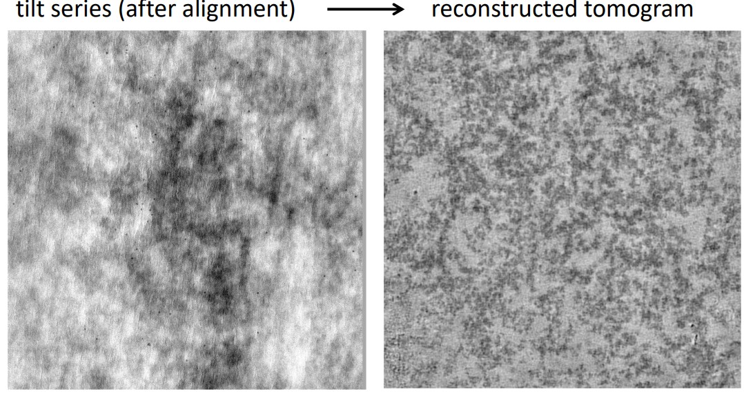
Figure 10.4: Reconstruction of a Tilt-Series
Fortunately, this problem can be overcome via multi-tilt tomography and sub-tomogram averaging.
10.1.1 Electron Tomography
3D electron tomography is used to analyze rare, large, and irregularly-shaped three-dimensional structures. It can be performed on isolated particles, macromolecular complexes, or on structures inside cells at room temperature or cryogenic conditions.
However, electron tomography is also limited by sample thickness (i.e., about 300 nm thick at most)4. It is also time-consuming and largely manual (in addition to being limited to small areas).
When performed under cryogenic conditions, the total electron dose that the sample can tolerate imposes a limitation (i.e., low electron dose results in a low signal-to-noise ratio).
As a general rule of thumb, 1 kV of acceleration voltage is needed to image 1 nm of a sample.↩︎