12.1 Nuclear Medicine
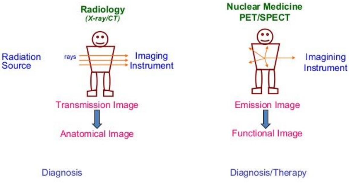
Figure 12.1: Graphical Illustration of Nuclear Medicine
Nuclear medicine is a specialty in medicine that uses radioactive substances in diagnosing and treating diseases. Approaches in this specialty record emitting radiation from within the body (instead of from external sources like X-rays).
Methods such as Single Photon Emission Computed Tomography (i.e., SPECT) and Positron Emission Tomography (i.e., PET) are two of the most common imaging modalities in nuclear medicine.
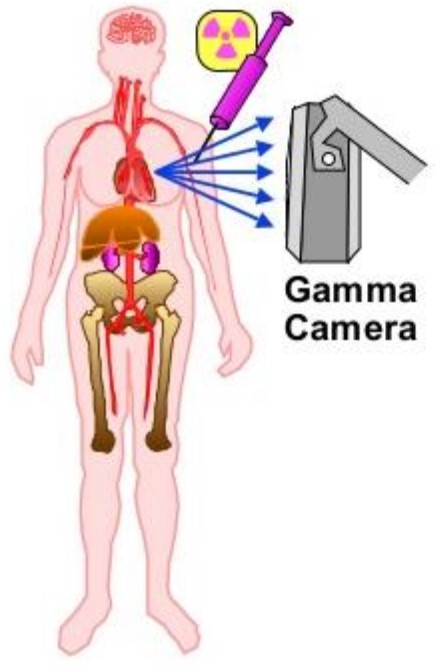
Figure 12.2: Workings of a Sample Nuclear Medicine Technique
For instance, when using radioactive substances in a patient, the following steps apply:
- The patient is injected with a small amount of a radioactive drug.
- The drug localizes in a specific location in a patient (depending on the nature of the drug itself).
- The drug radiates gamma radiation as it decays.
- The gamma rays that leave the patient are imaged.
12.1.1 Principles
Nuclear medicine provides a non-invasive method of determining physiological processes. They then localize in a specific area of the body (depending on their structures) and can provide information in the form of:
- Images
- Numerical data
- Time-activity curves
Depending on how nuclear medicine is used, it can be used to yield information about processes such as:
- Glucose metabolism
- Blood volume, flow, and perfusion
- Tissue and organ uptake
- Receptor binding and oxygen utilization
12.1.2 Gamma Cameras
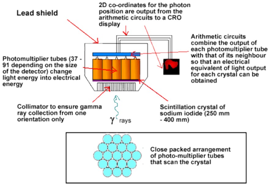
Figure 12.3: Workings of a Gamma Camera
A gamma camera uses gamma rays to image samples. A typical gamma camera has three parts:
- Collimators
- Scintillation detector
- Electronics and computer components
The camera has a large area of sodium iodide crystals with a lead collimator - this allows radiation to be transmitted from a particular point.
The above device can be moved to cover a larger area.
12.1.3 Radiopharmaceuticals
Radiopharmaceuticals are radioactive materials that are given to patients and consist of:
- A chemical molecule that determines how the radiochemical behaves once in vivo.
- A radionuclide. The radiation emitted from this molecule can be detected on the outside via an imaging device (e.g., a gamma camera) or a sample of body fluid (e.g., urine or plasma).
Diagnostic radiopharmaceuticals must deliver the minimum required dose of radiation while still obtaining the required information.
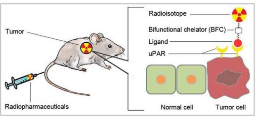
Figure 12.4: A Sample Radiopharmaceutical
Therapeutic radiopharmaceuticals must deliver the maximum required dose of radiation to the diseased organ or tumor while minimizing the amount of radiation bought to non-target tissues (e.g., bone marrow).
12.1.3.1 Ideal Radiopharmaceuticals
For in vivo imaging, the radiopharmaceutical should:
- Easily emit photons in high abundances in energies that can be picked up by a gamma camera (i.e., 25 keV to -300 keV).
- Not emit charged particles as these can be absorbed within a few millimeters of tissue. These greatly increase the amount of radiation that the patient gets.
- Have a short half life to keep the amount of radiation that the patient receives as low as possible.
For therapeutic purposes, the radiopharmaceutical should:
- Emit charged particles. These are typically beta particles, but could also be Auger electrons, internal conversion electrons, or even alpha particles.
- Have a low abundance of gamma photons - this allows the activity distribution to be imaged.
- Have a short half life (i.e., typically within several days).
12.1.3.2 Administering and Metabolizing Radiopharmaceuticals
Radiopharmaceuticals can be administered via the following methods:
- Intravenously or arterially
- Inhaled as a gas or as an aerosol
- Injested as a gas or as a solid meal
How a radiopharmaceutical is metabolized ultimately depends on its chemical properties - for instance:
- Some radiopharmaceuticals are simple ions - for example, sodium-131 iodide.
- Some radiopharmaceuticals are aggregates of molecules that are labelled with a radionuclide.
- Some radiopharmaceuticals are radio-labelled blood cells (i.e., red or white blood cells).
- Remainder radiopharmaceuticals are complex molecules such as phosphonates, peptides, and antibodies.
There are also two kinds of radiotherapies to be aware of:
Unsealed source radiotherapy
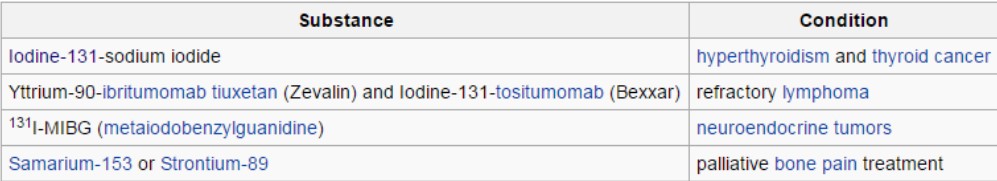
Figure 12.5: Commonly Used Nuclear Medicines for Unsealed Sources
This is the use of soluble forms of radioactive substances (that are typically ingested by the body via injection or ingestion).
Substances in this source are typically used for their Biological properties (which are similar to their non-radioactive parent substance).
Sealed source radiotherapy
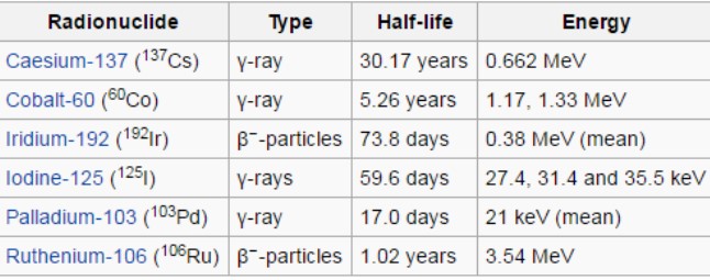
Figure 12.6: Commonly Used Radionuclides for Brachytherapy
The radioisotope remains in a capsule or in a metal wire during therapy and is not allowed to dissolve in fluids. This is typically referred to as brachytherapy.