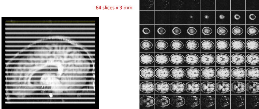
14.4 fMRI Image Acquisition
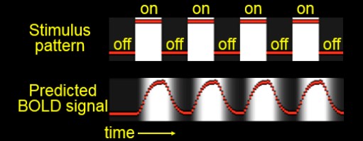
Figure 14.6: Stimuli in a fMRI Setup
The subject in question is given some sort of stimulus or task with a rest condition in between.
The image time series must then be analyzed to see where the signals change in response to stimulation.
fMRI image acquisition is rapid - it takes an image once per few seconds (i.e., 2 - 4 seconds). Anatomical images may take minutes to acquire.
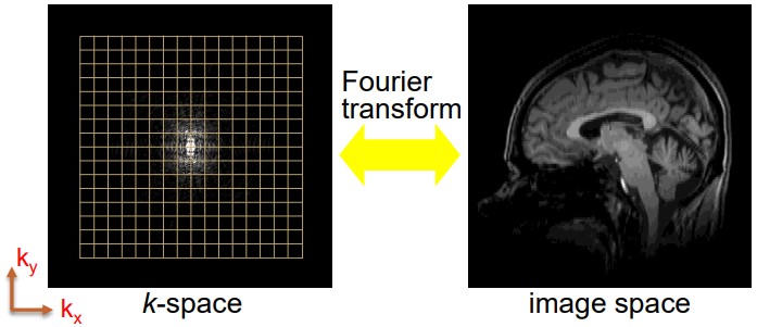
Figure 14.7: From a k-space to an image
The data in a k-space then undergoes a Fourier transform to yield the transformed image.
14.4.1 Shot Trajectories
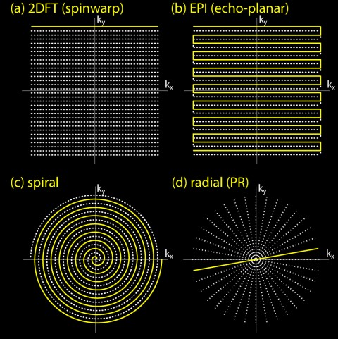
Figure 14.8: Trajectories in a fMRI Image Acquisition
The spiral fMRI is the only serious alternative to the EPI shown above. It has a short, apparent TE, is fast, and efficiently utilizes gradient hardware.
It also has different artefacts from a EPI.
14.4.1.1 Trajectory Considerations
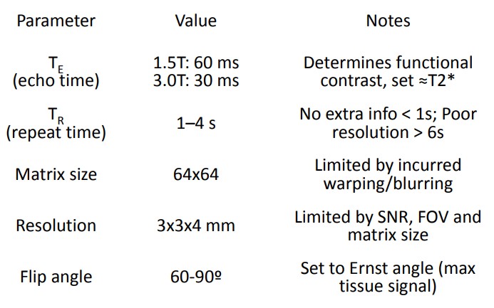
Figure 14.9: Parameters in a fMRI Image Acquisition
There are three considerations:
- A longer readout means more artefacts. For instance, single-shot (i.e., EPI and spiral) have warping and blurring.
- Cartesian vs. radial coordinates.
- SNR for N shots with time per shot Tread.
14.4.2 Acquisition Stages
There are two main kinds:
14.4.3 Limitations in Image Acquisition
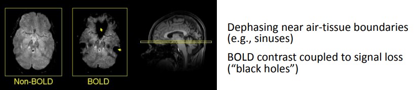
Figure 14.12: Dephasing in air-tissue boundaries
There are several:
BOLD Effect
BOLD contrasts are based on signal dephasing and requite a long delay for contrasts.
Signal Dropout in BOLD
Dephasing also occurs near air-tissue boundaries due to abrupt shifts in magnetic susceptibilties.
Image warping
Imperfections in the magnetic field, signals from readouts, and longer readouts can lead to the final image being altered.
Field offset
This introduces phase accrual during readouts. An EPI offsets warp images while a spiral offsets blur images.
Physiological noises
Physiological noises can be “BOLD-like” and increases with TE and B0.