2.2 Early Microscopists and their Microscopes
Robert Hooke and Antoine van Leeuwenhoek were two early microscopists whose works with the microscope profoundly contributed to modern Biology.
2.2.1 Robert Hooke (1653 - 1703)
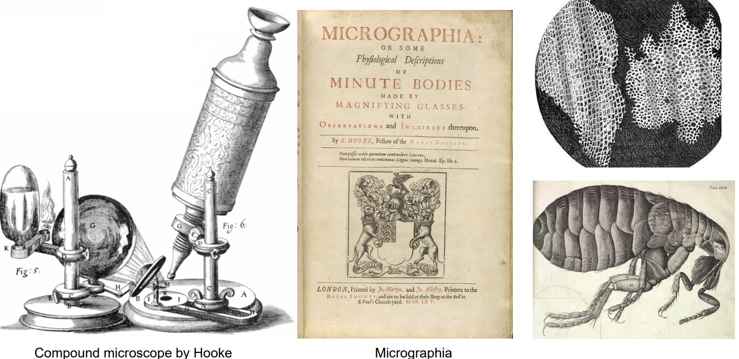
Figure 2.4: (from left to right) Robert’s Compound Microscope, his Published Work, and some of his Sketches.
He was a Dutch scientist whose work with the compound microscope led to the discovery of Cell Biology.
2.2.2 Antoine van Leeuwenhoek (1632 - 1723)
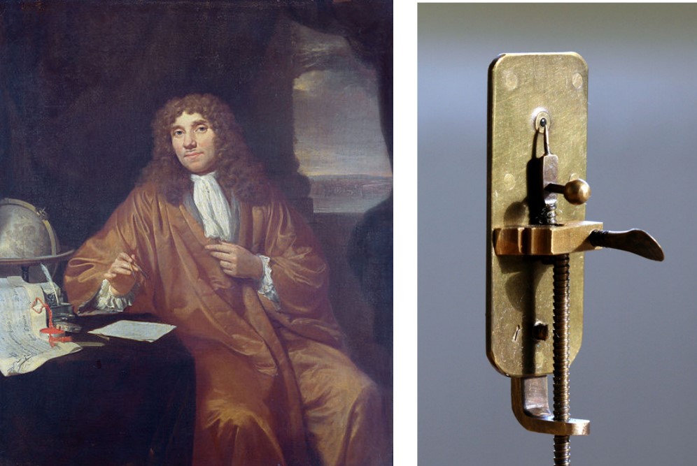
Figure 2.5: A Painting of Antoine and the Microscope that he Used.
Leeuwenhoek was a Dutch draper (i.e., cloth importer) able to identify the following organisms via his simple microscope. He carried a magnifying glass to inspect the quality of cloths (as part of his job):
- Protists
- Bacteria
- Sperm
- Vacuoles (i.e., an organelle)
Leeuwenhoek had no formal training in Science - however, he was still able to make a microscope via his passion (albeit a little small - see above graphic).
2.2.3 Comparing Hooke’s and Leeuwenhoek’s Microscopes
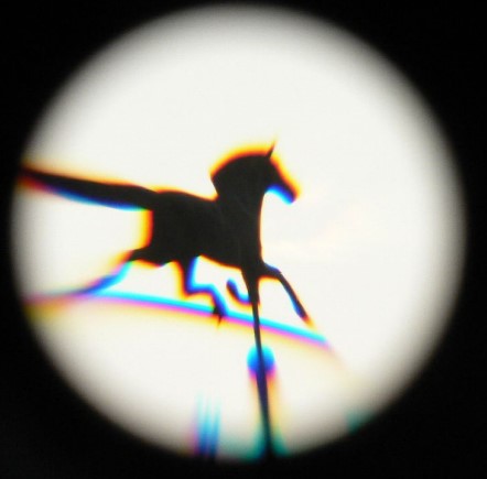
Figure 2.6: Chromatic and Spherical Aberrations of a Wind Vane through a Telescope
The following table notes some differences between Hooke’s and Leeuwenhoek’s microscopes. The above image also shows how an object may appear through a lens when it is affected by chromatic and spherical aberrations.
| Scientist | Difference |
|---|---|
| Hooke | Had chromatic and spherical aberrations |
| Leeuwenhoek | Superior to Hooke’s compound microscope |
An abberation (in the context of microscopy) is an imperfection in the final image seen by the viewer.
A spherical abberation happens when the light rays through a convex lens do not all converge to a single focal point. Consequently, the final image viewed is blurry (i.e., like a low-quality camera trying to zoom in - see the picture of the wind vane above).
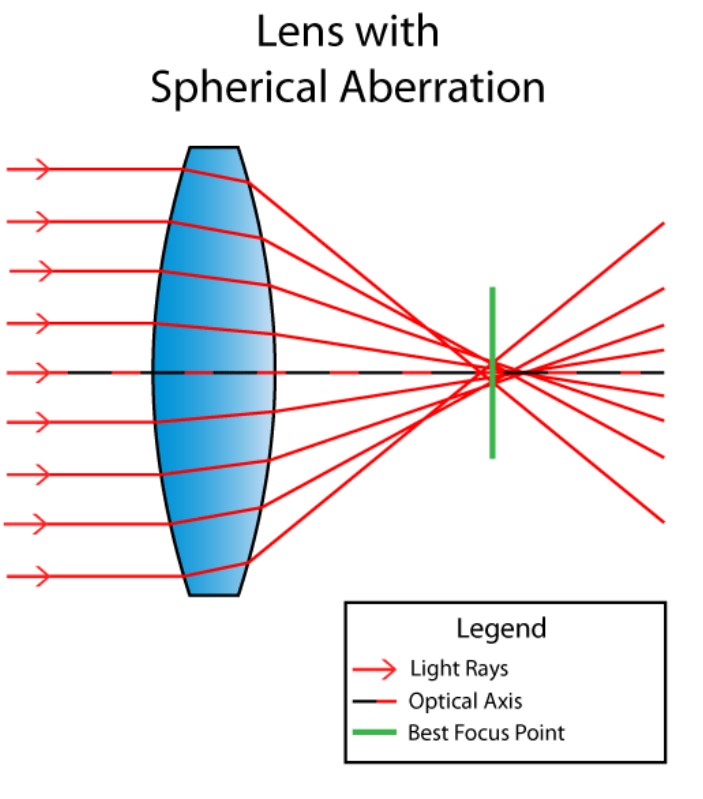
Figure 2.7: Demonstration of Spherical Abberation
A chromatic abberation is an imperfection that happens when the outline of an object becomes colored (see above image of wind vane again).