Topic 8 Basic Image Analysis
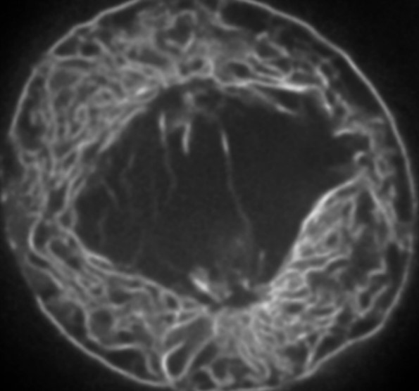
Figure 8.1: A Live HeLa Cell Visualized Using Confocal Microscopy
Modern microscopes, especially confocal ones, can generate thin optical section images of samples.
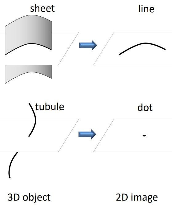
Figure 8.2: 2D Representation of 3D Structures
In a thin 2D cross-section, a 3D sheet could appear as a line while a 3D line could appear as a dot when viewed in 2D. Nevertheless, it is unreliable to deduce a sample’s 3D structure from their 2D images.