13.2 Performing MRI

Figure 13.6: Procedure for Taking an MRI
The following steps are typically done to obtain a MRI:
- Put the subject in a big magnetic field
- Transmit radio waves into the subject (usually between two to ten milliseconds)
- Turn off the radio wave transmitter in 2.
- Receive the radio waves that have been re-transmitted by the subject
- Convert the the radiofrequency data (i.e., RF) to an image
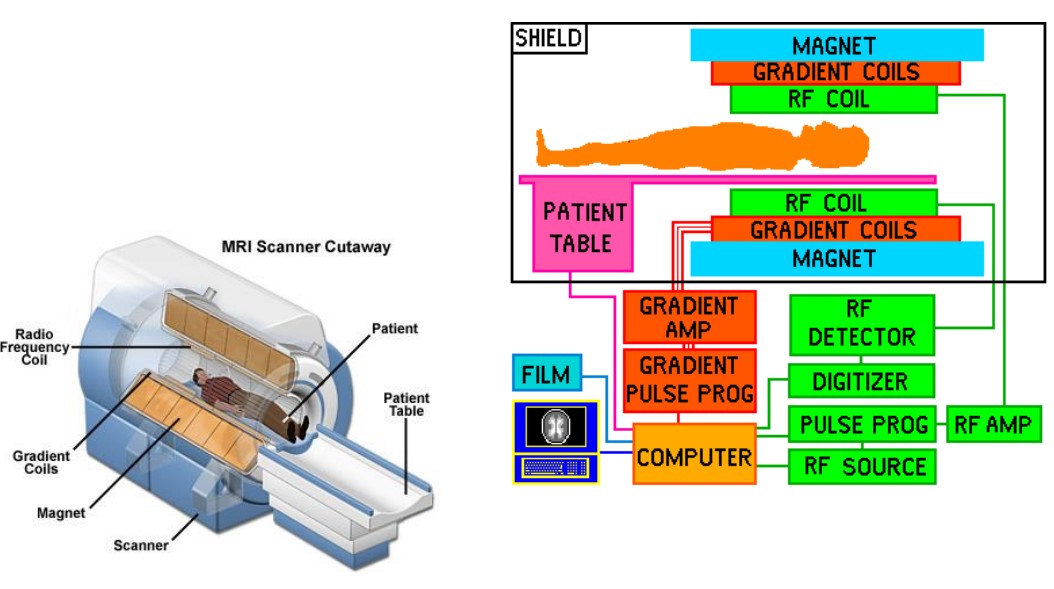
Figure 13.7: Schematic for an MRI Machine
A typical MRI machine has the following components:
- Static magnetic field coils
- Gradient magnetic field coils
- Magnetic shim coils
- Radiofrequency coils
- Subsystem control computer

Figure 13.8: Different Components of an MRI Machine
Components for data transfer, physiological monitoring, stimulus display, and behavioral recording hardware are also included.
13.2.1 Factors for Influencing MRI Quality
Many factors also influence the quality of a MRI image, including but not limited to:
- Quantum properties of nuclear spins
- RF excitation properties
- Tissue relaxation properties
- Magnetic field strength and gradients
- Timing of gradients, RF pulses, and signal detection.
13.2.2 Kinds of Nuclei for NMR
A nucleus has two peroperties: spin and charge. All nuclei are made up of protons and neutrons, both of which has 12 spin; protons are positively charged while neutrons are uncharged.
Opposing spins cancel each other out so that only atoms with an odd amount of protons or neutrons have spins.
As hydrogen is the most abundant atom in the body (i.e., most Biological tissues are predominantly carbon, hydrogen, oxygen, and nitrogen, and most of the body’s water is hydrogen) and is also the most sensitive to magnetic resonance imaging (i.e., MR), this makes it an ideal atom choice for performing MRI scans.
13.2.3 Use Cases for MRI
Physicians use MRIs to help diagnose or monitor treatments for conditions such as:
- Tumors
- Heart problems
- Blockages or enlargements of blood vessels
- Liver diseases (e.g., cirrhosis) and that or abdominal organs
- Diseases of the small intestine, colon, and the rectum.
However, MRI is ideally suited for soft tissue problems - for instance:
- Brain tumors
- Spinal infections
- Diagnosing multiple sclerosis
- Visualizing torn ligaments
- Visualizing shoulder injuries
- Evaluating bone tumors and slipped disks
- Diagnosing strokes
13.2.4 Disadvantages of MRI
Metal objects must be kept out of the room where the machine is operating; people with pacemakers cannot be safely scanned.
Some people also feel claustrophobic and may feel confined while undergoing an MRI procedure. Some people are also too big or fat to fit inside the MRI machine. Rapidly switching the magnetic field on and off can also cause nerve stimulation.
The machine makes a loud noise (i.e., up to 120 dB - equivalent to a jet engine’s takeoff) and also requires that people lay very still (for up to 90 minutes in some cases).
MRI systems are also expensive.