3.3 Phase Contrast Microscopy
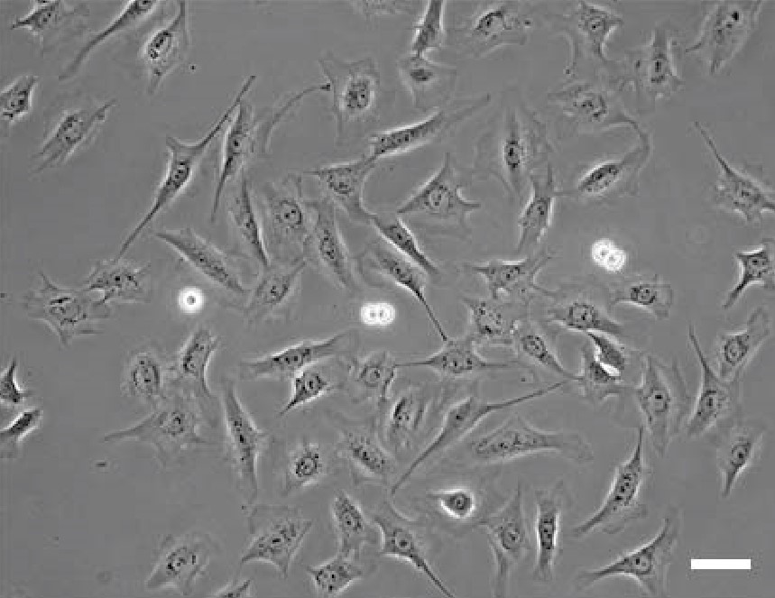
Figure 3.10: Phase Contrast Microscopy of Rat1 Cells
No prior staining is required to see cells.
A special kind of objective is also required - the objective phase ring must match the condenser phase ring (e.g., both Ph 2)
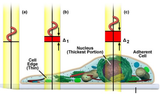
Figure 3.11: Different Refractive Indices of Organelles
The idea behind this is that different organelles have different refractive indices which results in different phase shifts. This translates to intensity differences in phase rings.