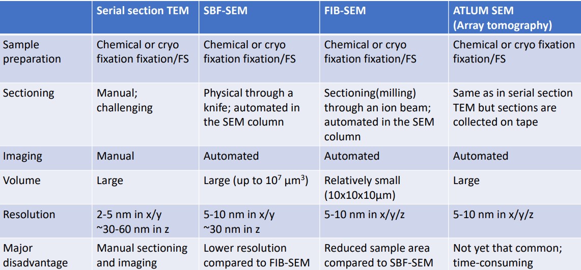
10.2 3D EM Volume Imaging
This part of the lecture focuses on the following two topics:
- Serial block face SEM (i.e., SBF-SEM)
- Focused ion beam SEM (i.e., FIB-SEM)
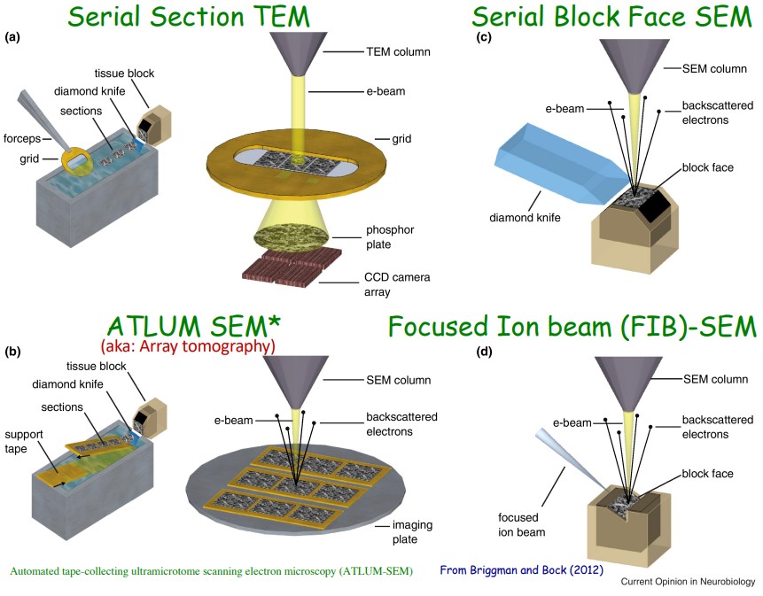
Figure 10.5: SBF-SEM and FIB-SEM Illustrations
10.2.1 Serial Section TEM / SEM
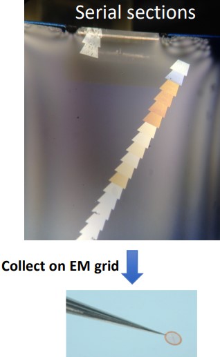
Figure 10.6: Manual Collection on EM Grids
60 to 80 nm sections are made for serial-section TEMs or serial section tomographies (i.e., thicker sections).
However, this is technically challenging - the order of the sections must be maintained. Losing sections loses information about the sample.
This process is also time-consuming as each section has to be individually imaged. The images must then be aligned and the area of interest reconstructed in 3D.
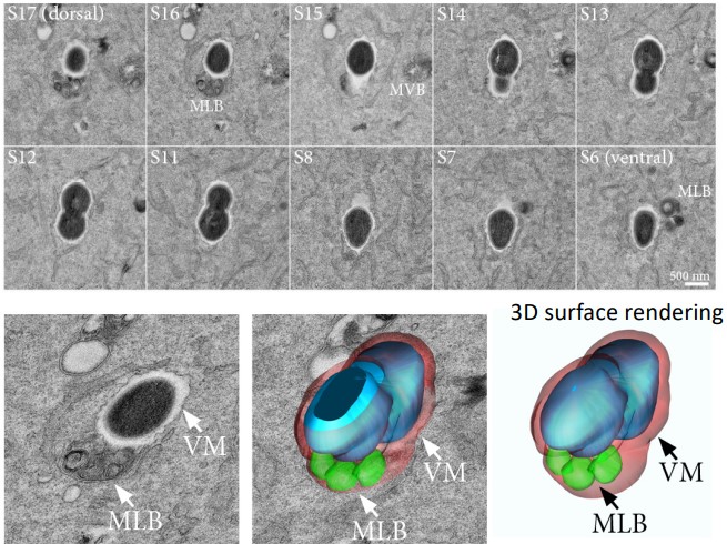
Figure 10.7: Serial Section TEM of Intracellular Bacteria
An array tomography can help automate the collection of sections on tape and image those sections in a SEM.