4.2 Excitation and Emission Spectra
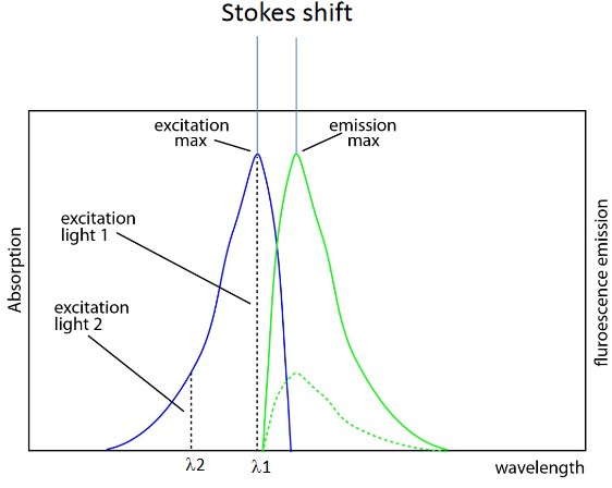
Figure 4.4: Emission and Absorption Spectra of Fluorescence Microscopy
Both spectra are normalized with the following properties:
Absorption spectrum
These plot absorption (i.e., y-axis) against wavelength (i.e., x-axis).
Emission spectrum
These plot flourescence intensity (i.e., y-axis) against wavelength (i.e., x-axis).
Emission spectra are independent of their absorption spectra due to the Stokes’ shift. Both spectra’s curves also appear like mirror images of one another.
4.2.1 Small Molecule Fluorophores
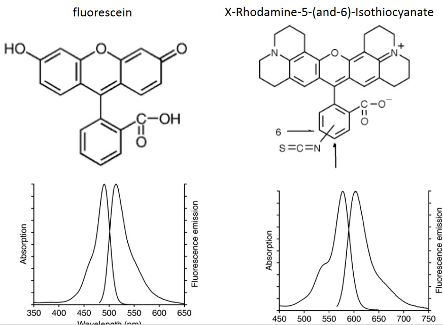
Figure 4.5: Examples of Fluorophores Used in Fluorescence Microscopy
Professor Lu Lei lists two examples of fluorophores and their emission and absorption spectrums.
4.2.1.1 Aequorea victoria’s GFP
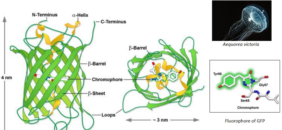
Figure 4.6: Tertiary Structure of a GFP
Note that GFP is short for Green Fluorescent Protein.
The GFP in this jellyfish (i.e., Aequorea victoria) can generate fluorescence on its own (i.e., no co-factors or enzymes are required). The fluorophore of the GFP is protected within the β-sheet barrel.
In Biology, GFPs are typically used to label a protein and is highly compatible with live cell imaging3.
4.2.2 Fluorescence Labelling Biological Samples
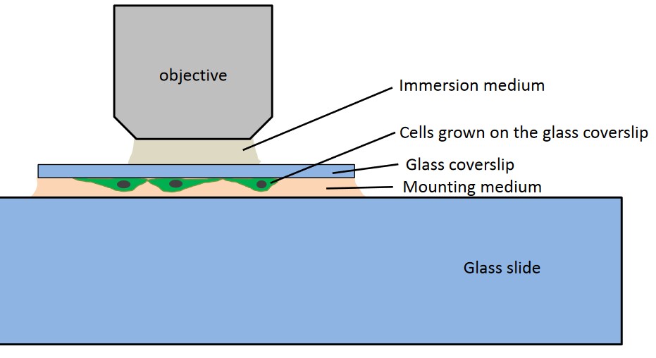
Figure 4.7: A Sample Immuno-Fluorescence Slide
After a cell is grown on a glass coverslip and fixated to it, the following steps can be performed:
Permeabilization
This allows the antibody to enter the cell. Detergent is used in this step to make “holes” in the cell.
Adding Primary Antibodies
These antibodies bind to a specific antigen and often sourced from mice, rabbits, and humans.
Washing
This removes unbounded antibodies.
Adding Secondary Antibodies
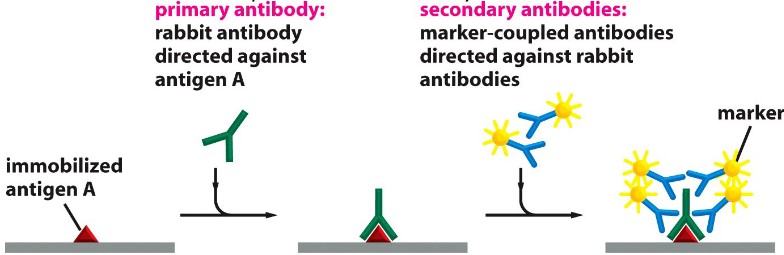
Figure 4.8: Primary and Secondary Antibody Labelling
These label the antibodies; goat antibodies conjugated with fluorophores are used here.
Washing
This serves the same purpose as before.
Mounting
This immobilizes the glass coverslip. The refractive index of the mounting medium should be the same as that of the immersion medium’s.
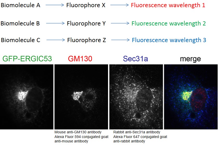
Figure 4.9: Multi-Color Labelling in Fluorescence Microscopy
With the above technique, multi-color labeling is also possible:
Though, note that many different fluorescence proteins are also available!↩︎