4.3 Wide-Field Microscopy
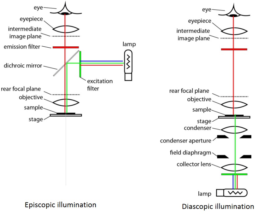
Figure 4.10: Different Kinds of Illuminations
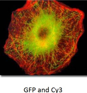
In wide-field epi-fluorescence microscopy, light that leaves the objective is excitation light. The objective acts as the condenser in this kind of microscopy.
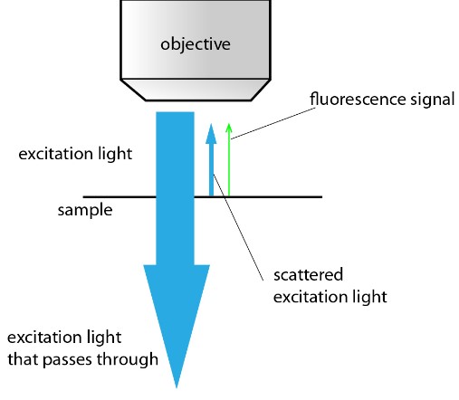
Figure 4.11: Passage of Light in an Epi-Fluorescence Microscopy
Light that enters the objective is fluorescence (which contributes to signals) and scattered emission light (which contributes to background signals).
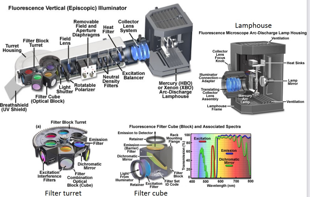
Figure 4.12: Optical Train of Epi-Fluorescence Microscopy
In this kind of microscopy, a high numerical aperture condenser acts as a condenser. This microscopy also has easy alignment and the sample could be mounted on a non-transparent support.
4.3.1 Filters in Epi-Fluorescence Microscopy
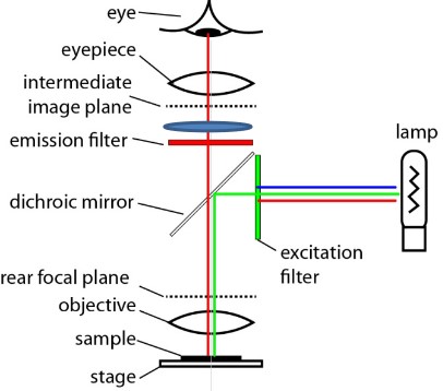
Figure 4.13: Filters in Epi-Fluorescence Microscopy
There are three main filters:
Excitation Filter
These select a bandwith of light to excite the fluorophore.
Dichroic Mirror
These reflect excitation light, transmit emission light, and suppress scattered excitation light.
Emission Filter
These pass fluorescence emission light and block scattered excitation light.
4.3.2 Kohler Illumination and Conjugate Planes
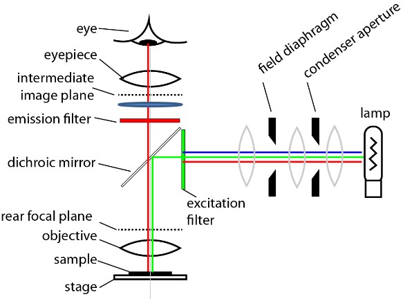
Figure 4.14: Planes in Epi-Fluoresence Microscopy
The planes are similar to bright-field microscopy. Kohler illumination is done when a sample is focused.
4.3.3 Resolution and Image Brightness
The resolution d of an epi-fluorescence microscope is:
d=0.61λNA
For a NA1.4 objective, d is usually about 200 nm.
The intensity is:
Intensity=NA4M2
Where M is the magnification of the objective.