12.2 SPECT

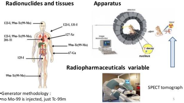
Figure 12.7: A SPECT Machine and its Results
Single Photon Emission Computed Tomography (i.e., SPECT) is a technique in nuclear medicine that uses gamma cameras to capture 3D information.
Information in a SPECT scan is shown as cross-sectional slices, but it can also be reformatted.
SPECT requires that a radionuclide is injected into the person (typically into the bloodstream).
12.2.1 How does SPECT Work?

Figure 12.8: A SPECT conducted with Tc-99m
Cameras in a SPECT scan acquire multiple, planar views of an organ’s radioactivity - these views are called projection views.
The views then get processed to create cross-sectional views of the organ (or the sample in question).
SPECT uses single photons made by gamma-emitting radionuclides, for instance, 99mTc, 67Ga, 111In, and 123I.
12.2.2 Comparing SPECT and PET Scans

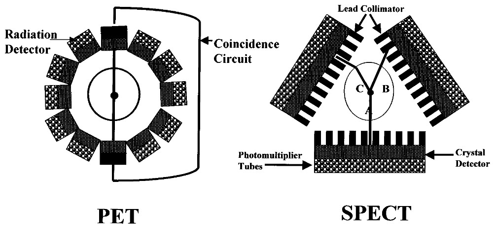
Figure 12.9: Setup of a PET and a SPECT machine
The table below lists some differences between a PET and a SPECT scan:
| SPECT | PET |
|---|---|
| Uses radiotracers | Uses radiotracers for positron decay |
| Captures photons in various directions | Decay makes two photons in opposite directions |
| Obtains projection images in various angles | Special circuitry used to detect two photons in opposing directions at the same time |
The above table lists some differences between a PET scan and a SPECT scan.
12.2.3 A Brief Overview on SPECT’s General Workings
The radionuclide in the victim’s body emits gamma radiation that are then picked up by a set of collimated radiation detectors.
Most detectors in current SPECT systems are based on single or multiple NaI(TI) scintillation detectors. Projection views are also acquired from around the victim.
